Scientific Calendar September 2025
Refractory anaemia with ring sideroblasts
How do ring sideroblasts store their iron?
Mitochondrial ferritin
Cytosolic ferritin
Transferrin complexes
Lysosomal granules
Congratulations!
That's the correct answer!
Sorry! That´s not completely correct!
Please try again
Sorry! That's not the correct answer!
Please try again
Notice
Please select at least one answer
Scientific background
Refractory anaemia with ring sideroblasts (RARS) is a subtype of myelodysplastic syndrome (MDS) characterised by anaemia and the presence of ring sideroblasts in the bone marrow [1]. In more recent classifications, this entity is referred to as MDS with ring sideroblasts (MDS-RS) and can be further subdivided into MDS-RS with single-lineage or multilineage dysplasia, based on the extent of bone marrow dysplasia [2]. RARS is defined by the World Health Organization as having 15% or more ring sideroblasts in the bone marrow [3]. Ring sideroblasts are erythroblasts that exhibit a perinuclear ring of at least five siderotic granules, covering at least one-third of the nuclear circumference [4]. In these cells, iron is stored as mitochondrial ferritin in the perinuclear mitochondria and can be detected with Prussian blue staining (Perls’ reaction) [3]. This histochemical staining technique is widely used for detecting iron in cells and tissues. It specifically identifies iron in the ferric (Fe³⁺) state, including forms bound in ferritin and haemosiderin [5]. Mitochondrial ferritin, present in RARS, is a ferroxidase enzyme that is not detectable in normal erythropoiesis [2].
Regardless of the underlying cause, the presence of ring sideroblasts typically indicates ineffective erythropoiesis and is observed exclusively in pathological conditions [6]. In contrast, normal bone marrow may contain ferritin sideroblasts, which should not be mistaken for ring sideroblasts. Ferritin sideroblasts are physiological erythroblasts that display a few blue granules scattered throughout the cytoplasm upon staining. These granules represent endosomes containing iron that has not been incorporated into haem synthesis [6].
RARS belongs to the clonal sideroblastic anaemias, whereas there are also non-clonal sideroblastic anaemias [2]. The course of RARS is typically characterised by an initial phase of erythroid hyperplasia and ineffective erythropoiesis, which can often remain stable for many years. In some patients, this may be followed by a phase of marrow failure, potentially leading to the later emergence of leukaemic blasts [6]. Therapy involves the prescription of erythropoiesis-stimulating agents (ESA), transfusion and iron chelation therapy [2]. The only curative treatment for MDS patients is allogeneic haematopoietic stem cell transplantation [2].
Case results
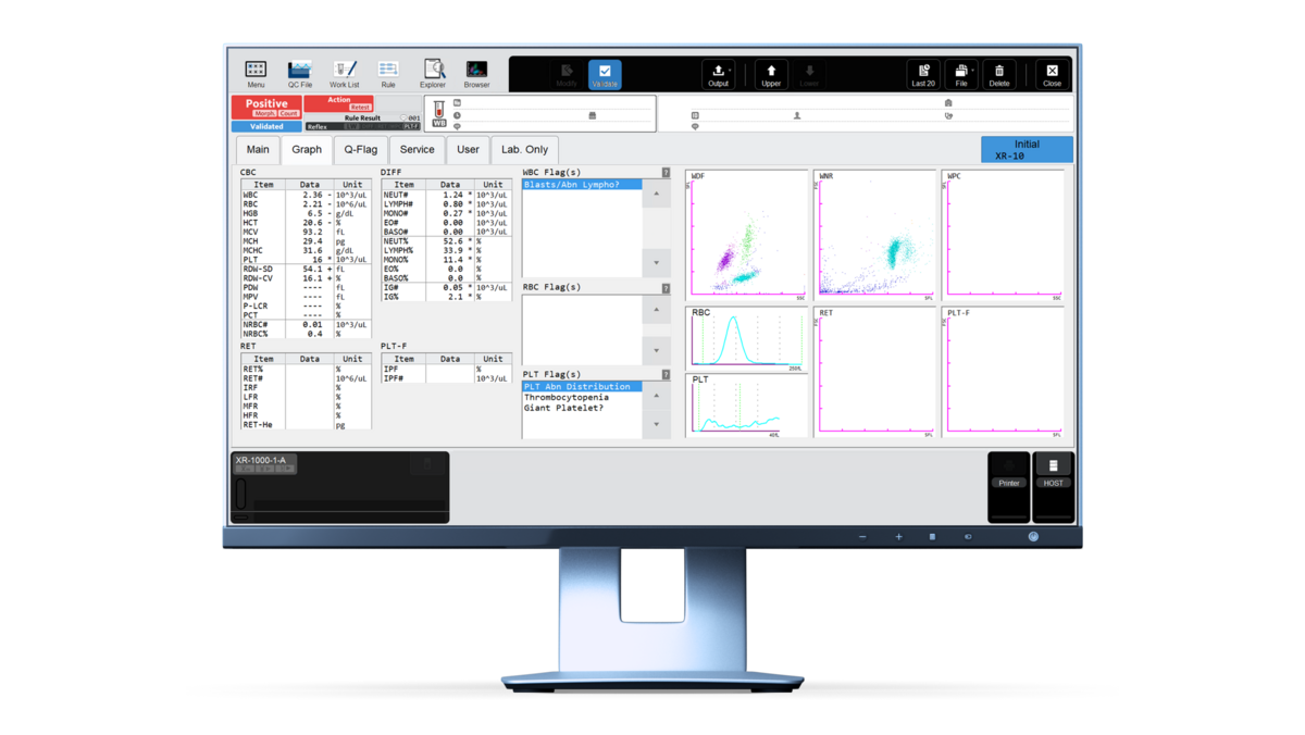
The blood count results of a referral patient revealed pancytopenia with a white blood cell (WBC) count of 2.36 × 10³/μL, a red blood cell (RBC) count of 2.21 × 10⁶/μL and a platelet (PLT) count of 16 × 103/μL. The haemoglobin value of 6.5 g/dL was also very low.

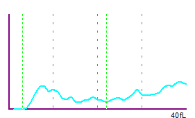
The platelet histogram showed an overall abnormal distribution curve, indicated by the flag ‘PLT Abn Distribution’. The platelet count was marked as unreliable. In this case, a reflex measurement to the PLT-F measurement channel was indicated by the analyser’s action message. The distribution of debris in the WNR measurement channel could have led the analyser to trigger the ‘Giant Platelet?’ flag. A follow-up of the platelet count is advisable.

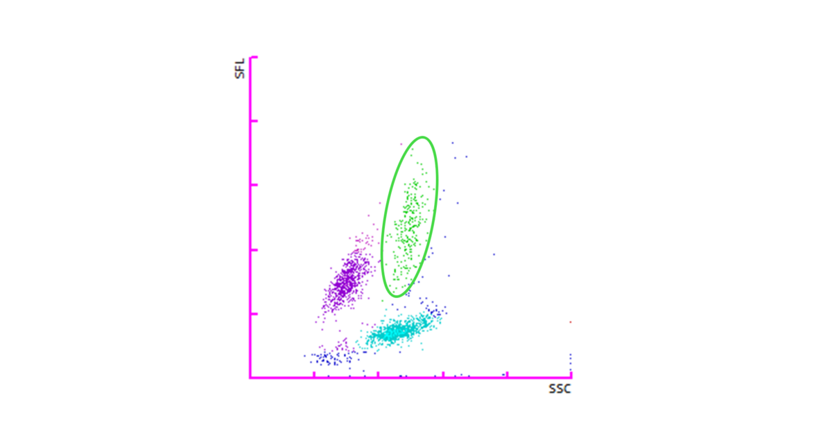
Despite the low WBC count, the automated differential appeared relatively evenly distributed, with only a slight upward trend of the monocyte cloud (green) in the WDF scattergram.

The analyser triggered the ‘Blasts/Abn Lympho?’ flag and a smear review was performed.
Due to the suspicion of an underlying malignant disease, a bone marrow aspirate was taken from the patient and investigated to confirm or rule out abnormal haematopoiesis.
Prussian blue staining (Perls’ reaction) of the bone marrow preparation exhibited erythroid precursors with a typical ring-shaped perinuclear pattern of blue iron aggregates (siderosomes). These cells are known as ring sideroblasts. The sum of all findings from the different laboratory investigations pointed towards the diagnosis of refractory anaemia with ring sideroblasts (RARS).
Downloads
References
[1] Arber DA et al. (2016): The 2016 revision to the World Health Organization classification of myeloid neoplasms and acute leukemia. Blood; 127(20):2391–2405.
[2] Patnaik MM et al. (2020): Refractory anemia with ring sideroblasts (RARS) and RARS with thrombocytosis: "2019 Update on Diagnosis, Risk-stratification, and Management". Am J Hematol; 94(4):475–488.
[3] Malcovati et al. (2013): Refractory anemia with ring sideroblasts. Best Pract Res Clin Haematol; (4):377–385.
[5] Meguro R et al. (2007): Nonheme-iron histochemistry for light and electron microscopy: a historical, theoretical and technical review. . Arch Histol Cytol; 70(1):1–19.
[6] Cazzola M et al. (2011): Ring sideroblasts and sideroblastic anemias. Haematologica; 96(6):789–792.


