
Cellules épithéliales
Les cellules épithéliales forment les épithéliums - qui constituent en quelque sorte les parois superficielles de notre corps. L'urothélium forme la paroi interne des voies urinaires, qui est un épithélium spécial construit par des cellules épithéliales. Chez un individu sain, un petit nombre de cellules épithéliales peut être présent dans l'urine. Toutefois, un nombre accru de cellules épithéliales dans l'urine peut être le signe d'une inflammation ou d'une infection, d'une infection des voies urinaires ou d'une contamination de l'échantillon.
Il existe trois types de cellules épithéliales que l'on peut trouver dans l'urine :
Les cellules épithéliales malpighiennes (Squa.EC)
Squamous epithelial cells are 20-100 µm in diameter, of multi-edged or rounded shape with a central or slightly off-centre nucleus, depending on their superficial, intermediate or deep layer position within the epithelium. Squa.EC form the urothelium of the urethra, vulva and reproductive system. The presence of Squa.EC in urine can indicate urethritis or injury through lithiasis or catheterisation. In many cases, it can be considered as a contamination.

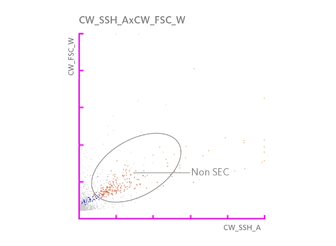
Non-squamous epithelial cells (Non SEC)
Non-squamous epithelial cells are 15-30 µm in diameter, of angular shape with a central or slightly off-centred nucleus. Non SEC cover the epithelium of the urethra, the prostate gland and uterine cervix. Non SEC can indicate urethritis and mechanical injury, but can also be a side effect of menstruation.
Transitional epithelial cells (Tran.EC)
Transitional epithelial cells are 15-150 µm in diameter, of polygonal, angular shape with a central or slightly off-centre nucleus, depending on their superficial, intermediate or deep layer position within the epithelium. Tran.EC form the multi-layered urothelium, covering the renal pelvis, kidney calyx, ureter, bladder and urethra. The presence of Tran.EC in urine can point to inflammation, malignancy, or injury through lithiasis or catheterisation.
Renal tubular epithelial cells (RTEC)
Renal tubular epithelial cells are 10-30 µm in diameter and appear in various shapes with an eccentric nucleus. RTECs comprise the monolayer epithelium, lining the proximal renal tubule, the loop of Henle, the distal renal tubule, the collecting ducts and the renal papilla. RTECs can indicate kidney damage or upper urinary tract infections in context of bacteriuria.
